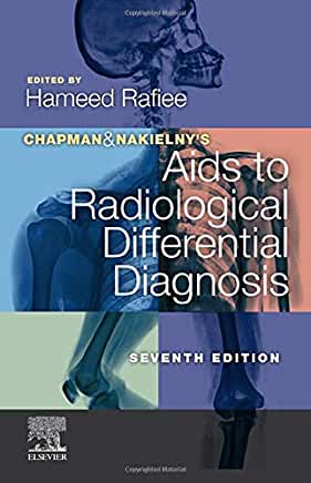
[This post is an update of my review of a previous edition of Aids to Radiological Differential Diagnosis, which I reviewed elsewhere in 2012. And granted, I won’t pretend the have done the kind of leg-work I did back then when I wrote, “I’ve been reading and annotating this book for close to a month now, and I barely scratched the surface in terms of absorbing the enormous amount of useful information stuffed into this relatively small 500 page handbook.” Still, I did have a close look at the new edition and will try to bring things up-to-date here.]
Aids to Radiological Differential Diagnosis (2020) is an extremely helpful handbook for interpreting imaging studies. For each radiographic finding, the book provides a differential diagnosis based on frequency, from most to least frequent. Take magacolon, for example, which is defined a colonic dilation greater than six centimeter. Radiologically this can be devided into two categories, toxic (with mural abnormalities) and non-toxic (without mural abnormalities.
The provided differential for toxic megacolon is:
- inflammatory bowel disease: ulcerative colitis, Crohn’s disease.
- Infection: especially pseudomembranous colitis.
- Ischemia (page 164).
On the other hand, differential diagnosis for non-toxic megacolon includes distal obstruction, paralytic ileum, chronic pseudo-obstruction, fecal impaction, Hirschsprung’s disease and hypoganglionosis, laxative abuse and amyloidosis.

This is exactly the kind of book you need to reach for when you see a radiographic finding, or read about one in a radiology report, and you want to explore alternative interpretations for those findings.
Perhaps even better, an entire section of the book, Part 2, is devoted to explaining key radiological findings, listed on the basis of the underlying diagnoses. This way, if you, as a clinician, already know—or think you know—the diagnosis you can look at this section to help you decide if the radiologic findings are compatible with the already-known diagnosis. For example, according to the authors (pages 617-8), hilar lymphadenopathy in patients with sarcoidosis should be bilateral and symmetric. Unilateral hilar adenopathy is uncommon and the same is true with regard to pleural involvement.
It would be very nice if future editions of this book showed simple schematics or drawings of the key radiographic findings. For example, I suppose that most radiologist know what “bone within a bone” appearance looks like, but it would be helpful for early learners to see a drawing of it while they are learning the relevant differential diagnosis (page 452). That way, they can read the book with needing to use a different resource to figure out what the pertinent findings actually are.
All in all, recommend Aids to Radiological Differential Diagnosis (2020) to anyone who reads and interprets imaging studies, especially general practitioners and radiologists. It is a terrific book to read and keep as a reference.


Leave a Reply