Medical Question: Flank Pain

A 60-year-old female presents to the emergency department complaining of intense left sided-flank pain radiating to the groin. Urinalysis is positive for gross hematuria, while a non-contrast CT of the abdomen and pelvis is negative for stones.

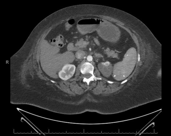
The patient is sent for a contrast CT of the abdomen and pelvic, from which the following image is taken:

Flank Pain and Hematuria


The patient was then re-examined at the bedside and her pulse was noted to be abnormal.

Question 1: What pulse abnormality would most likely account for the finding in the above image and why?

Question 2: What serum laboratory abnormality, if strongly positive, would best support this patient’s CT-evidenced diagnosis?

(Question 1 already answered and awarded per below. The Medical Media Review will give a $100 Amazon.com gift card to whoever posts the correct answer to Question 2 first. Offer expires August 31, 2015. See Contest Rules for additional details.)


Posted

in

, ,

by

Comments

5 responses to “Medical Question: Flank Pain”

  1. James (pseudonym) Avatar
    James (pseudonym)

    1. Irregularly irregular, which is a sign of underlying atrial fibrillation. the AF has lead to thromboembolic phenomenon and caused renal artery occlusion (?)

    2. Hyperkalemia?

  2. James (pseudonym) Avatar
    James (pseudonym)

    2. *hypokalemia

    1. Mark Yoffe, MD Avatar

      Congratulations, “James”!

      You’ve answering Question #1 correctly and precisely.

      On physical examination, an irregularly irregular rhythm was, in fact, identified in this patient. Further, an ECG confirmed atrial fibrillation. The contrast CT (shown) confirmed the diagnosis a left-sided acute renal infarction, likely secondary to a thromboembolus. (An alternative cause of this patient’s renal infarction might have been an abdominal aortic dissection. However, that would not be associated with any particular heart rhythm abnormality. In addition, an acute thrombus is a more common cause of an isolated, unilateral renal infarction, compared with an aortic dissection. In fact, as expected, this patient’s contrast CT was negative for aortic dissection.)

      I’ve emailed you at the address supplied to help you claim your prize. I will need you actual name and the email where you want the prize sent. If you don’t see the email, please check your spam folder because that’s where a lot of “Congratulations! You are the winner” emails end up!

      Unfortunately, I cannot give you credit for your answer to Question #2. This patient’s right kidney is enhancing beautifully – and is functional. Therefore, there is no reason for the patient to have hyperkalemia. (As an aside, there is no specific reason for the patient to have hypokalemia either. Granted, she has atrial fibrillation, but there are many potential causes for that).

      1. elham raofi Avatar
        elham raofi

        LDH and CRP is my answer to 2th question

        1. Mark Yoffe, MD Avatar

          One of these is correct. Which?

Leave a Reply

Your email address will not be published. Required fields are marked *