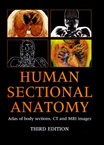
Human Sectional Anatomy: Atlas of body sections, CT and MRI images (2009) by Professor Harold Ellis is an exceptional anatomy book on many different levels. First, it shows you cadaveric sections that have been dissected in three dimensions to highlight some of the three dimensional sections of (what should have been) a two dimensional cut. It’s pretty hard to describe in words but of you look at the sections on the cover, for example, this becomes clearer:

These dissected cuts help the reader translate the two dimensional anatomy into a better understanding of three dimensional anatomy and of axial, sagittal and coronal CTs and MRIs.
Second, the book focuses heavily on clinically relevant anatomy. For example, the authors note:
Note the close relationship between the pons, medulla oblongata and the atlanto-occipital joints and dens of the axis (odontoid peg). This explains why injuries at the C1/C2 level are so serious and diseases that affect this region, e.g. rheumatoid arthritis … can be so disabling.
(Id. page 59)
The book is peppered with hundreds of similar clinical pearls.
Third, there are CT and MRI slices throughout the book which correspond, roughly at times, to the cadaveric sections. These images are also labeled for pertinent and clinically relevant anatomical features as well.
In future editions, I would like to see bigger pages. (The copy I purchased is subtitled “Pocket Atlas of Body Sections.” It is the size of an iPad, and so it is neither a handbook nor a full-sized atlas….)
I recommend this book very enthusiastically to medical student, residents, and attendings.

Leave a Reply